0:00
Hi friends, in this video let us
0:02
understand how to identify different
0:04
types of plant cells or tissues under
0:06
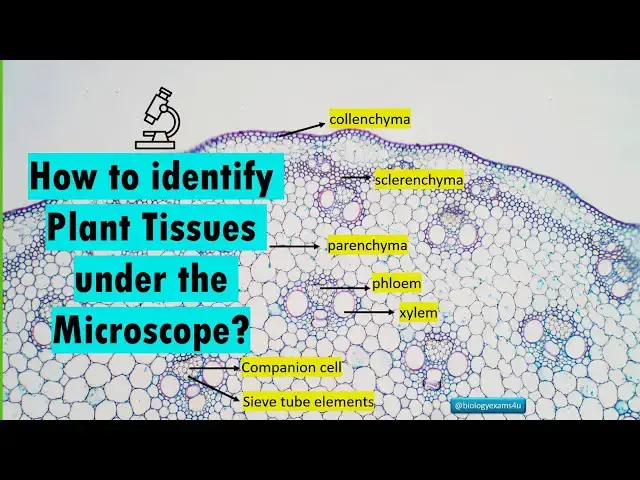
the microscope. So this is the
0:08
cross-section of a monocode stem under
0:10
the microscope. As you see there are
0:12
different tissues like colona then
0:15
scleranca, parenma, phium, syylum and
0:19
phium has companion cells and sift tube
0:22
elements. Once we have stained and
0:25
placed under the microscope, how to
0:27
identify these different types of cells
0:30
and let us see. So these are all tissues
0:33
we know. Paranky, scleranca, colonima.
0:36
Then the two conductive tissues asylum
0:38
and phm they are called permanent
0:40
tissues. Why they are called permanent
0:42
tissues? Because they lacks the ability
0:45
to divide and they have differentiated
0:47
after maturation. They have different
0:50
roles like protection, conduction,
0:52
storage etc. Permanent tissues are
0:55
classified into two. The first one is
0:57
the simple permanent tissue that is
0:59
composed of only one cell type like
1:01
parenma colanka and scleranca. The
1:04
second one is the complex permanent
1:06
tissue that is composed of many cell
1:08
types. Sylm has straits vessels parangma
1:12
etc. So it is made up of many types of
1:15
cells that makes it complex. Now let us
1:18
identify these tissues under the
1:19
microscope. The first is the paranka.
1:22
Paranka is most common tissue once we
1:25
have a section under the microscope. The
1:28
most common tissue we see is a paranka.
1:30
As you see here, these are all parang
1:33
cells. These cells are thin bold cells
1:36
without lignification
1:39
often equally sized cells with
1:41
intercellular spaces. They are living
1:43
cells at maturity. The staining pattern
1:47
is normally we use saffronine or fast
1:50
screen for staining. As these are thin
1:52
wall cells, these cells stain lightly
1:55
with saffron or fast screen. It has a
1:58
clear appearance often polygonal cells
2:00
with distinct but thin boundaries as you
2:03
see here. Primary function is storage.
2:06
Some paragamea cells are involved in
2:08
photosynthesis with chloroplast called
2:10
as chloroga. Then some are involved in
2:13
secretions secretreting various
2:14
substances like oils and parangma plays
2:18
a role in wound healing and
2:20
regeneration. And finally these are the
2:24
majority of cells that is within a
2:26
plant. So this maintains the torture of
2:29
cells and tissues making the system
2:31
tortured. The second type of tissue is
2:34
colanga. Cola as you see it is seen just
2:37
below the epidermis. Let me zoom in.
2:40
This is olamata cells. So as you see
2:43
these have thickened corners. There is
2:46
ligignification and uneven thickening as
2:48
you see in the cells often especially at
2:52
the corners. So these are living
2:53
mechanical tissue with an uneven
2:55
thickening in the primary cell walls
2:57
especially at the corners. Cells are
2:59
packed without intracellular spaces. It
3:02
provides flexible support to growing
3:04
organs. The most important thing is this
3:07
is a mechanical tissue, a living
3:08
mechanical tissue that of offers
3:10
flexibility or elasticity allows the
3:13
bending and swaying of plant parts
3:15
without breaking and that is offered by
3:18
this columatus tissue. The staining
3:21
pattern is this cells deeply stain with
3:24
saffroninine at corners or at regions
3:27
where there is thickening. Uh so
3:30
elongated it cells with distinct often
3:33
brightly stained thickened corners as
3:35
you see here. And the third one is
3:37
scaranga. Scarma as you see here this is
3:43
This is a dead mechanical tissue with
3:46
highly lignified walls. This has high
3:49
lignified secondary walls without
3:52
protolast provides structural support
3:55
and protection. There are two main types
3:58
of scaranga. First one is fibers that
4:00
are elongated pointed cells. The second
4:02
one is clear with irregularly shaped
4:05
cells often found singly or in groups
4:08
also called as stone cell. function is
4:11
mechanical support staining pattern. As
4:14
these cells have highly thickened
4:17
lignified secondary walls, these cells
4:19
stain deeply bright red due to the
4:22
presence of lignen. So easily visible
4:25
under the microscope when stained with
4:28
saffroninine. The next group of tissue
4:30
is the syllem. So let me zoom in. This
4:33
is a sylum vessel. These cells are
4:35
responsible for conduction of water
4:37
throughout the plant and also dissert
4:40
minerals from root to leaves. It is
4:42
composed of four main components. First
4:44
one is a traits elongated tapering dead
4:47
cells with ligignified walls and pits.
4:50
Then this is a vessels shorter wider
4:53
dead cells that is arranged end to end
4:56
forming a continuous tube with
4:58
perforation plates. And third one is
5:01
parangma. And this is a living cell that
5:04
is involved in storage
5:06
of food and lateral conduction of water
5:09
and finally syllem fibers which is
5:11
sclerank cells that provides mechanical
5:14
support to this vascular system or
5:16
syllem components. The staining pattern
5:19
is as these are highly liignified cells.
5:23
The struct and vessels stain brightly
5:26
red as you see here. Bright red walls of
5:30
these cells are prominent because of
5:32
linification. Silent paragma just like
5:34
ordinary paragma will stain lightly with
5:38
saffronine or fast green. Vessels and
5:40
traces appear brightly stained red tubes
5:44
as you see here with distinct wall
5:47
thickenings throughout. Whereas paranga
5:50
is seen just like ordinary parangata
5:53
cells with light staining. Functions
5:55
include water conduction primarily by
5:57
trucks and vessels then mineral
5:59
transport and also provide mechanical
6:02
support by means of xyllem fibers. And
6:06
the final tissue is the phium. As you
6:09
see this is the fium and this is a
6:12
vascular bundle. So let me zoom in. As
6:14
you see this is the foam
6:17
and this is a seaf tube element with a
6:20
white lumen or white cavity and the
6:22
smaller cell is a companion cell. The
6:25
sift tube element is without nucleus and
6:28
companion cell has nucleus and this
6:30
companion cells regulates the activity
6:32
or flow of uh sift tube safe tube
6:36
elements. So this is the diagrammatic
6:39
image. As you see this is there is a
6:40
safe plate and this is a safe tube and
6:43
close to the safe tube there is a cell
6:45
which is companion cell that is
6:46
nucleated whereas safe tube is without
6:50
nucleus and there are flu paranka also
6:53
and these are the function. The primary
6:54
function is food transllocation that is
6:57
from the site of synthesis which is a
6:58
source or leaves to the site of storage
7:01
which is called as a sink. composed of
7:03
four main elements. Zip tubes that is
7:06
living elongated cells that lack nucleus
7:08
at maturity. Then companion cells living
7:11
with nucleus that controls the activity
7:14
of Z tube. Then there is flamagma living
7:16
cells involved in storage and lateral
7:18
conduction and also phm fibers which is
7:21
also called as bas fibers that are
7:23
sclerang cells providing mechanical
7:26
support. The function is food
7:28
transllocation. Then there is storage by
7:30
fium parangma and also support
7:32
mechanical support provided by phium
7:36
cells present in the phium staining
7:40
lighter shade of saffronine fast green
7:43
compared to syllem. As you see here
7:45
these are thin wall cells. Floy fibers
7:48
as they are scenagamata cells will stain
7:50
brightly with zapronine due to lignen.
7:54
Overall appearance is zip tubes appear
7:56
as relatively wide whereas companion
7:58
cells are smaller and that is closely
8:01
associated with seep tubes and both are
8:03
likely stained as the walls are not
8:06
liignified or thickened. So let me
8:09
summarize. So these are the tissues we
8:11
identified under the microscope. The
8:13
first one is a colonima that is present
8:16
just below the epidermis. Then there is
8:19
clearanca. As you see here, it has
8:21
ligignified fold. So stain deeply red
8:24
with saffronin. Then barna lightly
8:28
stained thin volt living cells. This is
8:31
followed by vascular bundle. As you see
8:33
this is a phium. Then sylum. Syllem is
8:36
sylum vessels and traces are linified
8:38
therefore stain deeply with saffron red
8:41
color. Whereas phium se tube and
8:45
companion cells are thinfold. So stain
8:47
lightly. So this this is a companion
8:49
cell and this is a sift tube element
8:51
which has a large lumen or cavity close
8:54
to the sift tube element that is a small
8:57
cell with nucleus that regulates the
8:59
activity of this sift tube element is
9:02
the companion cell. Hope you can
9:04
identify the plant tissues under the
9:06
microscope. Take care stay blessed.
9:09
Thank you so much. You are the biologics