How DNA is packaged in eukaryotes? The ultimate packaging phenomenon
Think of a situation where approximately 2 meters of DNA is to
be packaged inside a cell which we cannot see. Rightly inside the nucleus of a
cell which is approx. 10μm in diameter, very much smaller than a cell. Wow! This
is the real situation. On an average, human cell contain 6.4 billion base pairs
of DNA distributed in 46 chromosomes.
- Distance between each base pair is 0.34 nm in length
- Then total length of DNA = 6x109 x 0.34x10-9=2.04 Meters
How this 2meters of DNA is packaged in a nucleus of approx 10μm?
How the stability of DNA is maintained in this mind boggling
packaging with accessibility to all enzymes and regulatory proteins required
for various processes like replication, transcription?
Many models were proposed and out of which two models stand
out.
Folded fibre model proposed
by E.J. Dupraw (1965)
- Chromosome consists of tightly folded fibre of 20-30nm diameter.
- Folded fibre consists of DNA histone helix of 3nm in a supercoiled condition.
- Histones were attached on the outside of the DNA coils that is histone shell around DNA
Nucleosome model
proposed by R.D. Kornberg (1974)
and
confirmed by P.Oudet (1975)
The best accepted model proposed for explaining this
ultimate packaging is ‘Nucleosome model’
or ‘Outdet concept of chromatin
structure’ or “beads on a string appearance” of chromatin under electron
microscope where DNA coils around histone proteins.
Before that let us discuss the major protein involved in
this packaging.
 |
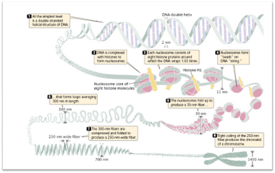
| DNA packaging at different levels |
Histones are the
major proteins involved in this packaging.
- The binding of the chromosomal DNA to histones is the first level of packaging.
- The DNA-histone protein complex is called the chromatin.
- There are 5 types of histones namely, H1, H2A, H2B, H3 and H4.
- These are basic proteins (25% of basic amino acids lysine and arginine) and negatively charged DNA (due to the presence of phosphate group) can easily bind to these positively charged proteins.
- Imagine a positively charged histone rod surrounded by negatively charged DNA thread.
- DNA: Histone ratio is 1:1.
Nucleosome model
- Nucleosome is the lowest level of chromosome organization.
- Nucleosome consists of a disc shaped structure of 11nm in diameter
- comprising of 2 parts: A core particle and a small spacer or linker DNA.
Core particle:
- consists of octamer of histones consisting of 2 copies of H2A, H2B, H3 and H4.
- A DNA strand of 146 bp is tightly wrapped around this core forming two circles (73 nucleotide bp/turn).
Spacer DNA or linker
DNA:
- length varies between species 1-80 bp
- One unit of H1 is associated with it.
- This makes the lowest level of chromosome organization and chromosomes are most condensed during metaphase.
A very very long rope of DNA packaged in a very very minute
nucleus of a cell. How? The answer is still largely unclear.
How many nucleosomes
are present in a human cell?
- Each nucleosome consists of 200 nucleotide pairs (approx)
- No. of nucleotide pairs in a human cell= 6x10⁹
- No. of nucleosomes= 6x10⁹/200=3x107 nucleosomes.
Tags:
‘Outdet concept of chromatin
chromatin
DNA packaging notes
E.J. Dupraw
Folded fibre model
H1
H2A
H2B
H3 and H4
Histones
linker DNA
Nucleosome model
P. Oudet
Spacer DNA
